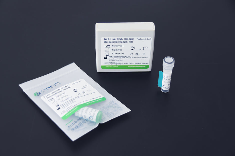
Approximately 300 million eligible women aged 35 to 64 in China need to undergo cervical cancer screening. From 2009 to 2017, the cumulative number of screened individuals was around 70 million, accounting for 23.33% of the actual screening population needed.
Faced with a large number of individuals requiring screening, accurate, simple, and efficient screening methods are particularly important. Currently, cytology combined with HR-HPV testing is the consensus for cervical cancer screening.
However, cytology screening faces limitations in sample collection, relies on the skills and experience of operators and readers, is subjective, has lower sensitivity, and is prone to missed diagnoses. HPV testing cannot differentiate between transient infections and transforming infections, and high rates of transient HPV positivity increase patient psychological stress.
Cytology-negative, HR-HPV-positive screening results are a common abnormality in clinical practice. A large number of referrals for colposcopy easily lead to overtreatment, and there is a lack of early risk prediction indicators for histological LSIL. Therefore, finding an objective biological indicator that can be used for triage management of cervical cancer and clarifying the risk of histological LSIL cervical lesions has important clinical value.
P16/Ki-67 Dual Staining in Cytology
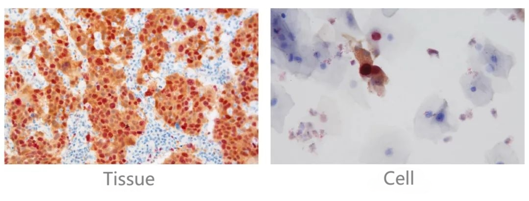
In normal cells, p16 is a marker of cellular quiescence, while Ki67 is a marker of cellular proliferation. These two markers are mutually exclusive and cannot be expressed simultaneously, much like a traffic light that cannot display both red and green at the same time. If they are co-expressed, it indicates a dysregulation of the cell cycle.
P16/Ki-67 dual staining in cytology is a valuable technique used to assess the cell cycle dynamics within tissues, particularly in the context of cervical cancer screening. By simultaneously visualizing p16, a marker of cellular quiescence, and Ki-67, a marker of cellular proliferation, this method provides crucial insights into the balance between cell growth and quiescence. In normal cells, the expression of p16 and Ki-67 is mutually exclusive, reflecting the well-regulated cell cycle. However, in dysplastic or neoplastic cells, the co-expression of p16 and Ki-67 indicates a disturbance in the normal cell cycle regulation, which can be indicative of precancerous or cancerous changes.
The utilization of p16/Ki-67 dual staining offers several advantages in the field of cytology and cervical cancer screening. Firstly, it addresses the limitations of HPV testing by providing enhanced specificity and sensitivity. Additionally, it reduces the subjective interpretation of results, as the assessment is primarily based on color determination, minimizing the impact of individual variability. Notably, the presence of p16/Ki-67 dual staining in even a single cell within a sample can be indicative of a positive result, enhancing the detection of abnormal cellular changes.
Furthermore, the application of p16/Ki-67 dual staining has been shown to effectively decrease the rate of missed diagnoses of high-grade cervical lesions, providing critical support for early intervention and treatment. This approach not only improves the accuracy of cervical disease detection but also minimizes unnecessary colposcopy examinations, leading to more efficient and targeted patient management. In summary, p16/Ki-67 dual staining in cytology represents a significant advancement in the field of cervical cancer screening, offering a more comprehensive and reliable method for identifying cellular abnormalities indicative of potential malignancy.
Advantages of p16/Ki-67 Dual Staining
Specificity and Sensitivity
The p16/Ki-67 dual staining technique in cytology offers significant advantages, including good specificity, which compensates for the limitations in specificity observed in HPV testing, and good sensitivity, which compensates for the lack of sensitivity in traditional cytology. This dual staining method addresses the shortcomings of both HPV testing and cytology, providing a more comprehensive and reliable approach to cervical cancer screening. By combining the strengths of both techniques, it enhances the accuracy of detecting cellular abnormalities associated with cervical cancer, ultimately improving the effectiveness of early detection and intervention.
Objectivity
The interpretation of results in p16/Ki-67 dual staining is solely based on color, making it impervious to subjective influences. This objective assessment reduces the potential for variability in interpretation, ensuring that results are consistently and accurately determined. As a result, the reliance on color-based determination enhances the reliability and robustness of the diagnostic process, contributing to more precise and dependable outcomes in the evaluation of cervical cytology.
Straightforward Criterion
In the context of p16/Ki-67 dual staining in cytology, the expression of both markers within a single cell in the sample is indicative of a positive result. This approach streamlines the interpretation process, allowing for a clear and unambiguous determination of positivity based on the presence of dual staining in individual cells. By establishing this straightforward criterion, the method simplifies result interpretation, contributing to greater efficiency and consistency in identifying abnormal cellular changes associated with cervical pathology.
Accuracy
This dual staining technique has demonstrated the capability to significantly decrease the incidence of missed diagnoses of high-grade cervical lesions. By improving the accuracy and sensitivity of cervical cancer screening, it plays a crucial role in identifying and capturing high-grade lesions at an earlier stage, thus facilitating timely intervention and treatment. This reduction in missed diagnoses contributes to enhanced patient outcomes and underscores the value of p16/Ki-67 dual staining as a powerful tool in cervical pathology assessment.
Making Prompt Therapeutic Measures Possible
By offering sufficient time and a solid foundation for clinical diagnosis and treatment, p16/Ki-67 dual staining contributes to the enhancement of early detection and intervention of cervical diseases. The comprehensive and reliable information provided by this technique enables healthcare professionals to make informed decisions regarding patient management, leading to more timely and targeted interventions. This proactive approach ultimately improves patient outcomes by facilitating early identification of cervical diseases and the prompt initiation of appropriate therapeutic measures.
Avoidance of Unnecessary Colposcopy Examinations
Relying on color interpretation results, p16/Ki-67 Dual Staining is not affected by subjective factors, making it widely used in cervical cancer screening. This helps to reduce the missed diagnosis and misdiagnosis caused by poor cytological sensitivity and HPV specificity, ultimately improving the detection rate of the disease. As a result, unnecessary colposcopy examinations can be reduced and avoided, leading to more efficient and accurate diagnosis.
Application in Various Scenarios
P16/Ki-67 dual staining is a valuable technique used in various scenarios. Firstly, it is utilized to identify samples that show negative results in cytology but are positive for high-risk HPV. This helps in identifying individuals who may require further evaluation or treatment. Secondly, it assists in identifying patients who test positive for high-risk HPV types other than HPV16/18, especially when HPV is used as the primary screening method. This ensures that individuals with a higher risk of developing cervical precancerous lesions are identified and managed appropriately. Additionally, p16/Ki-67 dual staining serves as an auxiliary diagnostic tool for cases with uncertain diagnoses, such as CIN2/ASC-H/CIN3 or below. It aids in providing more accurate and reliable results, reducing the chances of missed diagnoses or unnecessary interventions. Moreover, this technique aids in the differentiation of various cell types, including atrophy, immature phosphorylation, and repair cells, enabling a more precise diagnosis. Lastly, it is also used for screening abnormal cells in pregnant women, ensuring early detection and appropriate management.
Overall, p16/Ki-67 dual staining plays a crucial role in improving the efficiency and accuracy of cervical cancer screening, reducing unnecessary colposcopy examinations, and enhancing patient care.
P16/Ki-67 Dual Staining Detection Kit of Celnovte
Introduction of P16/Ki-67 Dual Staining Detection Kit
Celnovte, established in 2010, is a high-tech enterprise specializing in precision diagnostic instruments and reagents for tumors. With a strong focus on research, development, production, and sales, Celnovte has positioned itself as a leading provider of comprehensive solutions for tumor pathology diagnosis products. The company boasts a state-of-the-art production, quality control, and research and development system that adheres to GMP requirements.
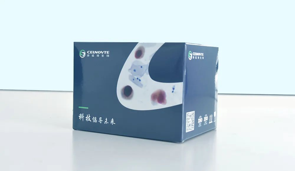
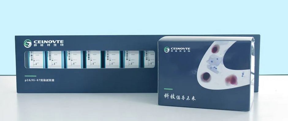
The P16/Ki-67 Dual Staining Detection Kit by Celnovte is a highly advanced diagnostic tool used for the detection of p16 and Ki67 biomarkers on cells or tissue samples. This innovative kit allows for the simultaneous detection of these biomarkers on a single slide, providing valuable assistance in the diagnosis of cervical precancerous lesions. The p16/Ki-67 cytological double staining technique, which relies on color interpretation results, is widely utilized in cervical cancer screening. It is particularly advantageous as it is not influenced by subjective factors, reducing the chances of missed diagnoses and misdiagnoses caused by poor cytological sensitivity and HPV specificity. By improving the disease detection rate, this kit plays a crucial role in enhancing the accuracy and efficiency of cervical cancer screening. Besides, Celnovte provides detection reagents with high-efficiency and high-accuracy of all kinds for diagnosis like Eosin Staining Solution and Progressive HE Staining Solution.
Components of P16/Ki-67 Dual Staining Detection Kit
P16/Ki-67 Dual Staining Detection Kit consists of components including peroxidase blocking agent, combined primary antibody, combined secondary antibody, fast red chromogen, activator, fast red buffer, DAB substrate, DAB buffer, and hematoxylin.
Results of Machine Detection

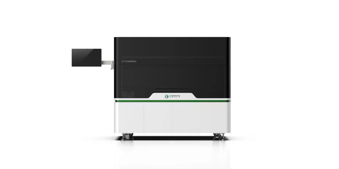
THE CNT 480 Fully Automatic Liquid-Based Cytology & Immunocytochemistry Stainer of Celnovte is an ideal instrument that can be used to gain the results of P16/Ki-67 dual staining efficiently and accurately.
Results of Manual Detection
