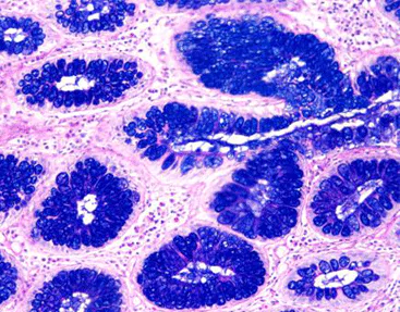
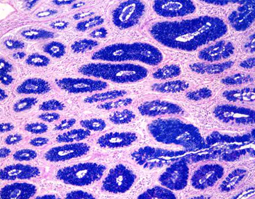
Alcian Blue and PAS stain techniques have long been pivotal in histological analysis, offering researchers valuable insights into the complex structures of tissues. The combination of these stains enhances the ability to evaluate various pathological conditions, particularly those involving mucinous substances. Innovations in the methodologies surrounding these stains continue to refine pathological diagnostics, ensuring higher accuracy and specificity. This article explores the latest developments in Alcian Blue and PAS combined staining techniques, emphasizing their significance in improving the accuracy of pathological diagnoses.
Components and Preparation of Alcian Blue and PAS Stain Kit
Alcian Blue Staining Kit
The Alcian Blue staining kit is integral to histological staining, particularly for visualizing acidic mucins in tissue samples. The staining process typically involves the use of Alcian Blue 8GX dye, which binds to carboxylated and sulfated mucopolysaccharides, highlighting areas rich in acidic mucins. Proper preparation and handling of the Alcian Blue stain are crucial; the dye is usually reconstituted in a sodium acetate buffer to maintain optimal staining pH. Furthermore, the quality and concentration of the Alcian Blue dye must be monitored carefully to avoid inconsistencies in staining results across different specimens.
Periodic Acid-Schiff (PAS)-Staining Technique
The PAS-staining technique serves as a complimentary method to Alcian Blue by highlighting neutral mucins and glycogen in tissue sections. This method involves oxidative treatment with periodic acid, which cleaves vicinal diols in carbohydrates, followed by the application of Schiff’s reagent. The resulting magenta staining provides contrast to the Alcian Blue results. Preparation for PAS staining also requires meticulous attention to detail, including the timing of each step and the condition of the reagents used. The PAS technique not only identifies neutral mucins but also plays a critical role in detecting fungal organisms and certain tumors, thus broadening its application in pathological evaluations.
Special Stain Techniques for Mucin Evaluation
Differentiating Neutral and Acidic Mucins
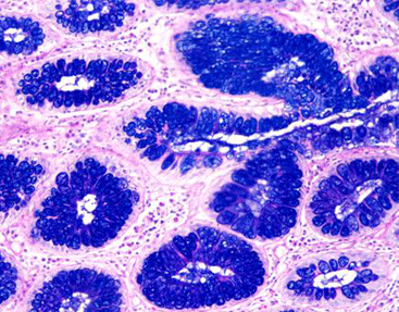
One of the pivotal advantages of employing both Alcian Blue and PAS stains lies in the ability to differentiate between acidic and neutral mucins. Alcian Blue demonstrates a strong affinity for acidic mucins, whereas PAS highlights neutral oligosaccharides, allowing pathologists to interpret mucin content accurately within tissues. This differentiation is particularly important in diagnosing conditions such as adenocarcinomas, where the expression of different mucin types can indicate the tumor’s origin and behavior. The ability to visualize and quantify these mucins through combined staining techniques provides a more comprehensive overview, improving diagnostic accuracy.
Historical Context of Special Stains
The development of special staining techniques, including Alcian Blue and PAS, has a rich history rooted in the evolution of histopathology. Early staining methods laid the groundwork for modern practices, utilizing basic dyes to enhance tissue visualization. As the understanding of mucins and glycoproteins proliferated, so did the sophistication of staining techniques. The innovation behind Alcian Blue and PAS has evolved in response to the clinical need for more precise diagnostic tools, reflecting advances in biochemistry and histology. This historical context underscores the importance of continual innovation in stain development to meet the demands of modern pathology.
Combining Alcian Blue and PAS Techniques
Methodology
The combined use of Alcian Blue and PAS techniques in histology represents a synergistic approach to tissue analysis. The methodology typically begins with the deparaffinization of tissue sections, followed by hydration. After applying Alcian Blue to visualize acidic mucins, thorough rinsing is essential to eliminate excess dye. The next step involves treating the sample with periodic acid, which sets the stage for the PAS reaction. Following this, Schiff’s reagent is applied, revealing areas containing neutral mucins. This sequential methodology not only optimizes staining efficiency but also enhances clarity and contrast in tissue samples, leading to a more reliable diagnostic outcome.
Enhanced Accuracy in Pathological Diagnosis
The integration of Alcian Blue and PAS staining techniques significantly improves the accuracy of pathological diagnoses. This combined approach allows for a comprehensive assessment of tissue characteristics, especially in tumors where mucin expression can signify critical information regarding tumor type and grade. Pathologists’ ability to differentiate between mucin types facilitates earlier detection of underlying malignancies and guides subsequent treatment decisions. Moreover, the dual staining enhances specificity, reducing the chances of misdiagnosis. Collectively, these advancements demonstrate how innovative staining techniques continue to evolve, providing invaluable tools for modern histopathology.
Celnovte Biotechnology Co., Ltd
Celnovte Biotechnology Co., Ltd, more commonly known as Celnovte, is a preeminent company dedicated to the research, development, production, and dissemination of cutting-edge pathological diagnostic reagents and instruments. Its forte lies in its comprehensive portfolio of Immunohistochemistry and in-situ hybridization solutions, which dominate the market.
Renowned for its exceptional MicroStacker™ IHC detection system, Celnovte boasts of unparalleled sensitivity and specificity, setting the industry benchmark. Additionally, the innovative PolyStacker™ technology has revolutionized the field by dramatically shrinking the processing time for frozen section IHC experiments to an impressive 10 minutes.
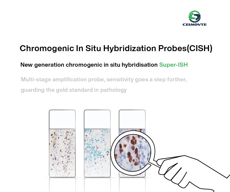
Moreover, Celnovte’s SuperISH™ RNA in-situ hybridization technology pioneers the detection of RNA targets with unparalleled precision, down to the single molecular level and single-cell resolution. The company also spearheads advancements in fully automated instrumentation, including IHC stainers, H&E stainers, specialized stainers, cytopathology instrumentation, and digital slide scanners, among others.
Contributions to Histology Staining Methods
Celnovte’s advancements in histology staining methods are particularly evident in its development of the Alcian Blue and PAS Stain Kit. This innovative kit streamlines the process of staining tissues, significantly reducing preparation time while ensuring consistent and reproducible results. The company has invested in research to optimize the formulation of Alcian Blue not only for better visual contrast but also for compatibility with a broader range of histological applications. Their ongoing research and development efforts address the evolving needs of pathologists, ensuring that staining methods remain aligned with contemporary diagnostic challenges.
Alcian Blue – PAS Stain Kit
Celnovte’s Alcian Blue – PAS Stain Kit, designed for specific staining procedures, comes in a convenient packaging of 4x10ml per kit, ensuring ample supply for various diagnostic needs. For optimal performance and preservation, it should be stored at a controlled temperature range of 2-8℃, ensuring that the reagents remain stable and effective over time. With a shelf life of 18 months, users can rely on the consistent quality and reliability of the kit for extended periods. By incorporating this stain kit into its extensive product portfolio, Celnovte further solidifies its position as a leading innovator in the industry, offering comprehensive solutions for a wide range of pathological diagnostic applications.
Future Prospects
Looking ahead, the future of Alcian Blue and PAS staining techniques is promising, with several avenues for enhancement and application. Continuous research into the biochemical interactions involved in mucin staining could lead to the development of even more sophisticated staining agents and methodologies. Innovations such as automated staining systems capable of performing Alcian Blue and PAS stains simultaneously may further streamline workflows in histopathology labs, thus improving efficiency while maintaining high standards of accuracy.
The integration of digital image analysis alongside traditional staining techniques may also play a crucial role in the future of diagnostic pathology. By employing advanced imaging technologies, pathologists can quantitatively analyze staining patterns, maximizing the diagnostic potential of Alcian Blue and PAS stains. This approach not only facilitates the early detection of malignancies but also aids in monitoring treatment responses by providing objective measurements of changes in mucin expression.
In conclusion, the combined techniques of Alcian Blue and PAS staining represent a significant advancement in histological diagnostics. As the field continues to evolve, the contributions of companies like Celnovte will be instrumental in fostering innovations that enhance the understanding and evaluation of tissue pathologies. The prospects for these advancements promise to not only refine diagnostic procedures but also improve patient outcomes in the ever-changing landscape of medical science.


