Multiplex Immunohistochemical (mIHC) Kits
What is Multiplex Immunohistochemistry?
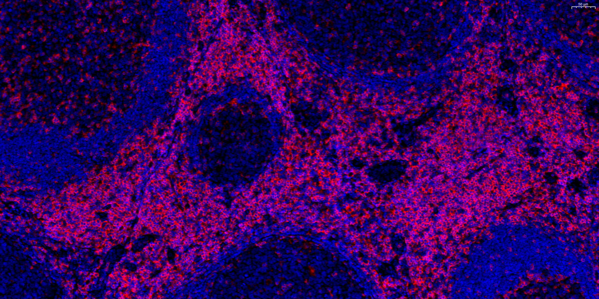
Multiplex Immunohistochemistry (mIHC) is an advanced technique that enables the simultaneous detection of multiple biomarkers within tissue samples. By utilizing specific antibodies that are tagged with different fluorescent dyes or chromogenic agents, mIHC allows for the visualization of multiple antigens in a single tissue section. This technique enhances the complexity of data that can be obtained in histological studies, improving the understanding of various diseases, especially in cancer research. The ability to observe interactions and spatial relationships between different cellular components provides more comprehensive insights than traditional single-stain immunohistochemistry.
Applications
The applications of the Multiplex Immunohistochemical (mIHC) Kit are numerous and span several fields, including oncology, immunology, and neurology. In cancer research, mIHC facilitates the identification of tumor microenvironments by allowing the simultaneous examination of markers that may indicate different cell types, such as immune cells infiltrating tumors. Additionally, mIHC has important applications in the study of autoimmune diseases, where it can be employed to dissect complex immune responses. Other areas of interest include neuroscience, where mIHC can help map neurodegenerative diseases by analyzing multiple proteins associated with neural functions.
The Technology Behind mIHC
High-Throughput Multiplex Staining
High-throughput multiplex staining technologies have revolutionized tissue analysis by providing an efficient means to pool data from multiple markers simultaneously. This approach not only saves time by reducing the number of slides that need to be processed but also conserves valuable tissue samples. The Multiplex Immunohistochemical (mIHC) Kit offers a combination of automation and advanced imaging techniques to streamline staining workflows, providing researchers with rapid access to results. Consequently, this high-throughput capability supports large-scale studies that demand robust data sets.
Automation and Faster Scanning
Automation in mIHC is essential for improving efficiency and reducing the potential for human error. Modern staining machines and imaging systems designed for mIHC allow for rapid and consistent processing of multiple samples. With the ability to complete staining operations in a shorter timeframe, researchers can expedite their analyses without compromising quality. These advancements in automation also facilitate the integration of image analysis techniques, leading to faster scanning and data extraction processes, further streamlining research workflows.
Advantages of Using mIHC Kits in Research
Cost-Effective Tissue Analysis
The implementation of Multiplex Immunohistochemical (mIHC) Kits can significantly reduce costs associated with tumor marker analysis. By enabling the simultaneous assessment of multiple biomarkers on a single slide, researchers conserve both tissue specimens and associated reagents. This approach minimizes the expenses incurred during typical sequential staining procedures. Hence, mIHC stands as a cost-effective alternative when evaluating complex tissue samples in research laboratories.
Highly Reproducible Results
Achieving highly reproducible results is crucial in scientific research. The application of the Multiplex Immunohistochemical (mIHC) Kit ensures that experiments maintain consistency due to its standardized protocols and associated controls. These features lead to reduced variability between experiments, enhancing the reliability of findings across different samples and studies. Consequently, researchers can confidently validate their results, uphold scientific integrity, and support more impactful conclusions.
Improved Data Accuracy
Utilizing mIHC leads to improved data accuracy by allowing simultaneous detection of multiple biomarkers in a single tissue section. This capability minimizes potential misinterpretations that can arise when analyzing each biomarker separately. The enhanced specificity provided by mIHC contributes to detailed insight into pathological processes and biomarker correlations. Consequently, researchers can make more informed decisions in their data analysis, resulting in breakthroughs and refined hypotheses in their studies.
Celnovte’s Multiplex Immunohistochemical (mIHC) Kit
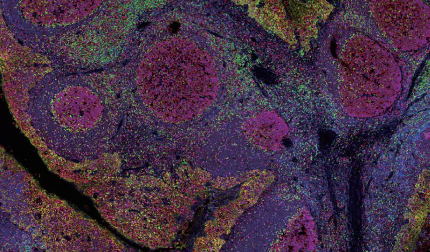
Celnovte‘s Multiplex Immunohistochemical (mIHC) Kit represents a significant advancement in the field of biological research, offering researchers the capability to detect up to 6-8 markers simultaneously in a single tissue sample. This groundbreaking technology eliminates the need for species restrictions on antibody selection, allowing for greater flexibility and versatility in experimental design. By simultaneously targeting multiple markers, the kit enables researchers to obtain a more comprehensive and nuanced understanding of biological processes within precious samples, thereby maximizing the value of these often limited resources.
Furthermore, the Celnovte mIHC Kit enhances the sensitivity of target detection and ensures the generation of specific image signals, providing researchers with clearer, more accurate results. This heightened precision is crucial for advancing scientific understanding and enabling the development of more effective treatments and therapies. Additionally, the kit’s automated detection system ensures high efficiency, stability, and repeatability, streamlining the research process and reducing the potential for human error. In summary, Celnovte’s Multiplex Immunohistochemical Kit represents a powerful tool for advancing the frontiers of biological research, enabling researchers to gain deeper insights into complex biological processes with unprecedented precision and efficiency.
Celnovte
About Celnovte
Celnovte is a leading biotechnology company dedicated to providing innovative solutions for researchers in the field of immunohistochemistry and tissue analysis. With a strong commitment to advancing scientific understanding, Celnovte specializes in the development and manufacturing of high-quality multiplex immunohistochemical (mIHC) kits that cater to a diverse range of applications. The company’s focus on research and development ensures that its products meet the needs of today’s scientists, facilitating enhanced research efficiency and data quality.
Developing Advanced mIHC Protocols
Enhancing 7-Color Panels with Multiple Biomarkers
One of the remarkable features of the Multiplex Immunohistochemical (mIHC) Kit is its capability to enhance 7-color panels that allow the simultaneous detection of multiple biomarkers in a single tissue section. This is accomplished by utilizing carefully selected antibodies tagged with distinct chromogenic or fluorescent labels. The ability to visualize multiple antigens concurrently enriches the amount of information derived from each sample, providing insights into the complex interactions within the tissue microenvironment. Such advancements are essential, particularly in oncology, where understanding tumor heterogeneity and microenvironmental influences can significantly impact diagnosis and treatment strategies.
Tyramide Signal Amplification Protocols
The Tyramide Signal Amplification (TSA) methods integrated into Celnovte’s mIHC Kit further improve sensitivity and signal detection. By harnessing the power of enzymatic amplification, TSA allows for minute levels of antigen detection that might otherwise go unnoticed in traditional staining techniques. This amplified signal is critical when dealing with weakly expressed biomarkers or rare cell populations within tissue samples. Ultimately, incorporating TSA protocols into mIHC workflows helps researchers obtain more precise results, leading to a deeper understanding of disease mechanisms, particularly in challenging cases where traditional methodologies may fall short.
Key Biomarkers: CD3, CD8, CD163, PD-L1, FoxP3, Cytokeratin (CK)
Celnovte’s mIHC Kit is designed to target a specific panel of key biomarkers that are pivotal in various research applications. For instance, CD3 and CD8 are critical in identifying T-cell populations that play significant roles in the immune response, while CD163 serves as a marker for alternatively activated macrophages, offering insights into the tumor microenvironment. PD-L1, a well-established immune checkpoint, is vital for evaluating immune response and therapy effectiveness in cancer patients. Additionally, biomarkers such as FoxP3 and Cytokeratin (CK) are instrumental in the study of immune regulation and epithelial status, respectively. Together, these biomarkers empower researchers to conduct comprehensive analyses and derive meaningful conclusions regarding disease processes.
Summary
The application of the Multiplex Immunohistochemical (mIHC) Kit not only enhances research efficiency but also provides high-quality, reproducible results that are crucial for advancing scientific understanding. As the demand for high-throughput, reliable, and informative research continues to grow, Celnovte remains at the forefront of innovation in the field of immunohistochemistry, enabling discoveries that contribute to improved diagnostics and therapeutic strategies.


